

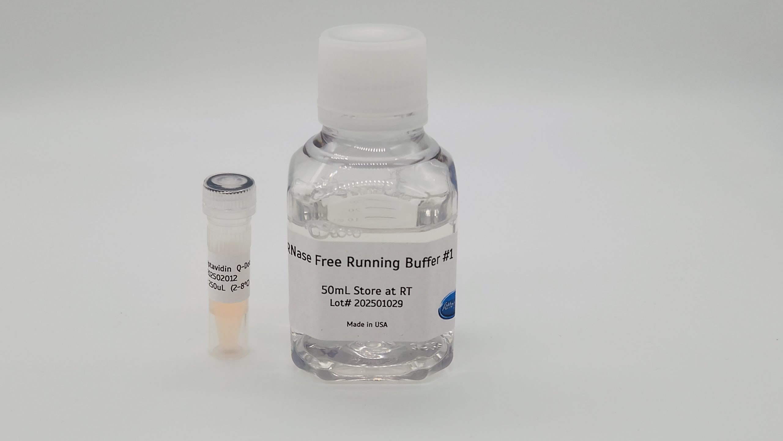


Streptavidin CdSe/ZnS Quantum Dots For Lateral Flow are ready-to-use streptavidin quantum dots for fluorescent lateral flow assays and other biotin-mediated detection systems. Each vial contains 250 µL of streptavidin-conjugated CdSe/ZnS core–shell quantum dots at 0.08 µM, with an emission peak of 655 nm, and the product ships with an RNase-free running buffer.
Streptavidin is covalently attached to the particle surface, and a proprietary surface coating supports colloidal stability and consistent fluorescence. Conjugates are functionally evaluated under lateral flow conditions. You can order the conjugate with either tetrameric or monomeric streptavidin to suit your assay architecture.
Key Features
- Bright red emission: CdSe/ZnS core–shell quantum dots with an emission peak of 655 ± 10 nm and a quantum yield above 50%.
- Covalent streptavidin coupling: streptavidin is covalently conjugated to the quantum dot surface for stable biotin capture.
- Two streptavidin formats: choose tetrameric or monomeric streptavidin at checkout.
- Ready to use: supplied at a working concentration to reduce preparation steps and run-to-run variability.
- Running buffer included: 50 mL of RNase-free Running Buffer #1 for fluorescent assay workflows.
- Tested format: functionally evaluated under lateral flow assay conditions.
Specifications
Each order of streptavidin quantum dots includes:
- Streptavidin quantum dot conjugate: 250 µL at 0.08 µM
- Running buffer: RNase-free Running Buffer #1, 50 mL, pH 7.6, 0.1% sodium azide, stored at room temperature
Conjugate specifications, per the lot specification sheet:
- Core/shell: CdSe/ZnS quantum dots
- Emission peak: 655 ± 10 nm
- Absorption: 440–650 nm
- Particle size: 9–11 nm
- Quantum yield: greater than 50%
- Solvent: 50 mM borate buffer, pH 8.3, with 0.05% sodium azide
- Storage: 2–8 °C, away from light. Do not freeze.
- Streptavidin format: tetrameric or monomeric
You will also need: your own biotin-labeled oligonucleotide or other biotinylated binder, lateral flow strips or another assay format, and a way to excite and read fluorescence. Custom concentrations, bulk volumes and alternative formats are available on request.

Who It’s For
These conjugates suit assay developers who want a fluorescent label instead of a colored one. Typical users include:
- Teams building fluorescent lateral flow assays for nucleic acid or protein targets that carry a biotin tag.
- Researchers who already use streptavidin gold and want to compare a fluorescent readout on the same assay design.
- Labs developing biotin-mediated detection systems that need a stable, ready-to-use streptavidin label.
How to Use Streptavidin Quantum Dots in Your Assay
The specification sheet gives this starting guide for biotin-labeled oligonucleotides:
- Bring the conjugate to room temperature and shake vigorously for about 30 seconds so the particles are fully dispersed.
- Add 100–150 µL of running buffer to your tube or well.
- Add 0.5–1 µL of the streptavidin quantum dot conjugate. If you dilute the particles, use 50 mM borate (boric acid) buffer, pH 8.3.
- Add 0.5–10 pmol of biotin-labeled oligonucleotide.
- Run the strip. Run times range from about 15 to 45 minutes.
To spray or spot the conjugate onto conjugate pads, contact sales@attogene.com for recommendations. Full details are in the streptavidin quantum dots specification sheet (PDF).
Tetrameric or Monomeric Streptavidin?
Streptavidin binds biotin with very high affinity, which is why it is a common bridge between a biotinylated probe and a detection label. Native streptavidin is a tetramer with four biotin-binding sites. Monomeric streptavidin has a single binding site, which some designs use to limit cross-linking when a target carries several biotin tags. The best choice depends on your assay architecture. If you are unsure, contact our team before ordering.
For a complete fluorescent starting point, see the Fluorescent Universal Lateral Flow Assay Kit (Quantum Dots). For a visual readout, compare Streptavidin Colloidal Gold for Lateral Flow. Other fluorescent labels include Streptavidin Europium Chelate Microspheres and Streptavidin R-Phycoerythrin.
Storage and Handling
- Store the conjugate at 2–8 °C away from light. Low-temperature storage helps preserve particle shape, size and stability.
- Do not freeze. Freezing causes irreversible aggregation and destroys the product.
- Particles may settle over time. Shake well before each use and check visually that all material has redispersed.
- If particulates remain, sonicate for 1 minute, shake and repeat as needed. Do not sonicate longer than 1 minute at a time to limit heating.
- Store the running buffer at room temperature.
Frequently Asked Questions
What is the emission wavelength of these streptavidin quantum dots?
The emission peak is 655 ± 10 nm, per the specification sheet. The listed absorption range is 440–650 nm.
What concentration and volume are supplied?
Each vial contains 250 µL of conjugate at 0.08 µM in 50 mM borate buffer, pH 8.3, with 0.05% sodium azide.
How much conjugate should I use per test?
The starting guide uses 0.5–1 µL of streptavidin quantum dots with 100–150 µL of running buffer and 0.5–10 pmol of biotin-labeled oligonucleotide. Optimize these amounts for your own assay.
Can I freeze the conjugate?
No. Freezing causes irreversible particle aggregation. Store at 2–8 °C away from light.
Can I get a different concentration or a bulk volume?
Yes. Custom concentrations, bulk volumes and alternative formats are available on request. Email sales@attogene.com.
For research use only. Not for use in diagnostic procedures. Questions? Call 512-333-1330 or email sales@attogene.com.


